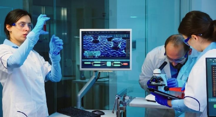
Importance Of Advanced Imaging Tools In Immunology Research
Immunology, the study of the immune system, has rapidly advanced due to the growing need for a deeper understanding of immune responses. As immunology specialists expand their knowledge, sophisticated tools are increasingly essential for analyzing immune mechanisms. Advanced imaging technologies are crucial in detailed insights into immune cell dynamics and interactions.
These cutting-edge techniques have revolutionized immunology research by enabling precise observation of immune cell behavior and their interactions with pathogens. This enhanced understanding has led to more effective diagnostics and treatments for immune-related disorders.
These imaging tools are significant, pushing research boundaries and driving breakthroughs that improve immunology diagnosis, treatment, and patient care.
Overview Of Different Types Of Advanced Imaging Tools
In immunology research, various advanced imaging tools have revolutionized the study of the immune system. These tools include:
- Fluorescence Microscopy: Visualizes specific molecules or cell types by labeling them with fluorescent probes, revealing immune cell dynamics such as migration and activation.
- Confocal Microscopy uses laser scanning to provide high-resolution, three-dimensional images of immune cells and tissues, capturing detailed structures and spatial relationships within complex environments.
- Super-Resolution Microscopy: Techniques like STED and PALM achieve resolutions beyond traditional light microscopy, allowing detailed visualization of subcellular structures and interactions between individual immune cells.
- Multiphoton Microscopy: Uses near-infrared light to penetrate deeper into tissues, enabling real-time observation of immune cell behavior in their natural environments.
- Imaging Flow Cytometry: Combines flow cytometry with high-resolution imaging to analyze immune cell phenotypes, morphologies, and functions at the single-cell level.
These advanced imaging tools have expanded researchers’ ability to explore the immune system’s complexities, leading to discoveries and insights.
Fluorescence Microscopy In Immunology Research

Fluorescence microscopy is a cornerstone of immunology research; key benefits:
- Multicolor Labeling: Simultaneously tracks various immune cells and their interactions, revealing complex immune responses.
- Spatial Organization: Visualizes immune cells within tissues, aiding in understanding the immune system’s structure and function.
- Dynamic Processes: Real-time imaging of cell migration, activation, and interactions, uncovering immune response mechanisms.
- Technological Advancements: Enhanced by sensitive probes and integration with super-resolution and multiphoton microscopy, driving research and treatment innovations.
This technique remains vital for advancing our understanding of the immune system and developing new treatments for immune-related disorders.
Confocal Microscopy In Immunology Research
Confocal microscopy has become a crucial tool in immunology, a detailed view of immune system structures and dynamics. Utilizing focused laser scanning and sophisticated optics, it provides high-resolution, three-dimensional (3D) images of biological samples, revealing immune cells’ spatial organization and interactions and their associated molecules.
Key Advantages:
- High-Resolution 3D Imaging: Confocal microscopy generates detailed, multilayered images by sequentially scanning samples and collecting light from specific focal planes. This has been instrumental in understanding the complex architecture of lymphoid organs, such as the spleen and lymph nodes.
- Subcellular Dynamics: Using fluorescent probes, researchers can visualize the organization and trafficking of cellular structures and signaling molecules. This detailed view is essential for studying immune cell activation and differentiation and understanding immune-related diseases.
- Intravital Imaging: Techniques like intravital imaging allow real-time observation of immune cells within living tissues, providing insights into their interactions with surrounding components such as blood vessels and extracellular matrix.
- Integration with Other Imaging Modalities: Developing faster scanning speeds, improved resolution, and integrating imaging techniques such as CT scans and MRIs enhance the capabilities of confocal microscopy. These advancements, supported by technologies from Tellica Imaging (https://tellicaimaging.com/), enable a more comprehensive analysis of immune system dynamics and more profound insights into immune cell interactions and mechanisms.
Confocal microscopy’s ability to deliver unprecedented insights into immune system structure and function makes it an indispensable tool in modern immunology. It drives research and leads to breakthroughs in diagnosing and treating immune-related disorders.
Super-Resolution Microscopy In Immunology Research
Super-resolution microscopy has revolutionized immunology by revealing nanoscale details of immune cells. Key techniques include:
- STED Microscopy: Provides high-resolution images of immune cell structures and molecular interactions beyond traditional light microscopy.
- PALM: Allows precise imaging of protein distribution and molecular assemblies in immune cells.
- Other Methods: Techniques like structured illumination microscopy (SIM) and expansion microscopy further enhance spatial resolution.
These advancements provide new insights into immune cell dynamics and function, advancing diagnostics and treatments for immune disorders.
Multiphoton Microscopy In Immunology Research
Multiphoton microscopy has become a vital tool in immunology with unique advantages for studying the immune system in living tissues:
- Deep Tissue Penetration: This technique uses near-infrared light to visualize immune cells within intact tissues, revealing their behavior in native environments.
- Real-Time Tracking: Enables dynamic observation of immune cell responses to stimuli, providing insights into activation and interactions.
- Clinical Applications: Helps visualize immune cell dynamics and tissue architecture in immune-related disorders, aiding diagnosis and monitoring.
These capabilities make multiphoton microscopy essential for advancing research and clinical practices in immunology.
Imaging Flow Cytometry In Immunology Research
Imaging flow cytometry combines the high-throughput analysis of flow cytometry with detailed microscopy imaging deep insights into immune cells.
Key Advantages:
- Detailed Analysis: Provides high-resolution images of individual cells, revealing phenotypes, morphologies, and functional properties.
- Rare Cell Identification: Detects and characterizes rare immune cell subsets and subtle changes linked to disease or therapy.
- Dynamic Interactions: Observe immune cell interactions within their microenvironment.
- Phagocytic Studies: Enhances understanding of phagocytosis in macrophages and dendritic cells.
Imaging flow cytometry continues to evolve, complementing techniques like CT scans and MRIs, driving advancements in immunology research and clinical practice.
Advancements And Future Prospects Of Advanced Imaging Tools In Immunology
The field of immunology has transformed significantly in recent years thanks to rapid advancements in imaging tools. These cutting-edge technologies have revolutionized how immunologists conduct research, enabling unprecedented insights into the system’s structures and dynamics. Advanced imaging techniques such as fluorescence microscopy, confocal microscopy, super-resolution microscopy, multiphoton microscopy, and imaging flow cytometry have all played crucial roles. These tools have greatly enhanced our understanding of immune responses and diseases by providing detailed views of immune cells, interactions, and environments.


